Image Gallery
Gallery
Localization of T. cruzi Aquaporin in Epimastigotes.

Media Details
Created 1/20/2004
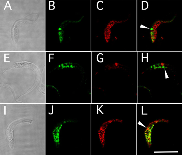
Localization of T. cruzi aquaporin in epimastigotes. (A-D) GFP-AQP expressing epimastigote co-labeled with anti-TcPPase: (B) GFP-AQP view, (C) TcPPase view. (E-H) GFP-AQP expressing epimastigote co-labeled with TRITC-concanavalin A to label the flagellar pocket: (B) GFP-AQP view, (C) concanavalin A view. (I-L) Wild-type epimastigote labeled with anti-TcPPase (J) and anti-TcAQP (K). Arrowheads mark an vacuolar (non-acidocalcisomal) localization of AQP near the flagellar pocket. Scale bar = 10 um.
Credits
- P. Rohloff , Department of Veterinary Medicine
- R. Docampo , Department of Veterinary Medicine